
Corneal Mapping
Corneal topography, also known as photokeratoscopy or videokeratography, is a non-invasive medical imaging technique for mapping the surface curvature of the cornea, the outer structure of the eye. Since the cornea is normally responsible for some 70% of the eye’s refractive power, its topography is of critical importance in determining the quality of vision.
The three-dimensional map is therefore a valuable aid to the examining ophthalmologist or optometrist and can assist in the diagnosis and treatment of a number of conditions; in planning refractive surgery such as LASIK and evaluation of its results; or in assessing the fit of contact lenses. A development of keratoscopy, corneal topography extends the measurement range from the four points a few millimeters apart that is offered by keratometry to a grid of thousands of points covering the entire cornea. The procedure is carried out in seconds and is completely painless.
Digital Retinal Imaging
Digital Retinal Imaging allows your eye doctor to evaluate the health of the back of your eye, the retina. It is used to detect diseases, such as, Diabetes and Macular Degeneration. It is critical to confirm the health of the retina, optic nerve and other retinal structures.
Many eye diseases, if detected at an early stage, can be treated successfully without total loss of vision. Your Retinal Images will be stored electronically. This gives the Doctor a permanent record of the condition and state of your retina. This is very important in assisting the Doctor to detect and measure any changes to your retina each time you get your eyes examined, as many eye conditions, such as Glaucoma are diagnosed by detecting changes over time.
OPTOS Retinal Exam
Annual eye exams are vital to maintaining your vision and overall health. We offer the optomap® Retinal Exam as an important part of our eye exams. The optomap® Retinal Exam produces an image that is as unique as your fingerprint and provides us with a wide view to look at the health of your retina. The retina is the part of your eye that captures the images you see, similar to film in a camera.
See Dr. Proske’s personal experience with the optomap® Retinal Exam, and how it saved his life.
Many eye problems can develop without you knowing. You may not even notice any change in your sight. But, diseases such as macular degeneration, glaucoma, retinal tears or detachments, and other health problems such as diabetes and high blood pressure can be seen and are often first detected with a thorough annual examination of the retina.
An optomap® Retinal Exam provides:
- A scan to show a healthy eye or detect disease.
- A view of the retina, giving your doctor a more detailed view than he/she can get by other means.
- The opportunity for you to view and discuss the optomap® image of your eye with your doctor at the time of your exam.
- A permanent record for your file, which allows us to view your images each year to look for changes.
The optomap® Retinal Exam is fast, easy, and comfortable for all ages. To have the exam, you simply look into the device one eye at a time and you will see a comfortable flash of light to let you know the image of your retina has been taken. The optomap® image is shown immediately on a computer screen so we can review it with you.
Please schedule your optomap® Retinal Exam today!
For more information on the optomap® Retinal Exam, go to the Optos website.
Visual Field Testing
A visual field test measures how much ‘side’ vision you have. It is a straightforward test, painless, and does not involve eye drops. Essentially lights are flashed on, and you have to press a button whenever you see the light. Your head is kept still and you have to place your chin on a chin rest. The lights are bright or dim at different stages of the test. Some of the flashes are purely to check you are concentrating. Each eye is tested separately and the entire test takes 15-45 minutes. Your optometrist may ask only for a driving licence visual field test, which takes 5-10 minutes. If you have just asked for a driving test or the clinic doctor advised you have one, you will be informed of the result by the clinic doctor, in writing, in a few weeks. Normally the test is carried out by a computerised machine, called a Humphrey. Occasionally the manual test has to be used, a Goldman. For each test you have to look at a central point then press a buzzer each time you see the light.
Optomap
The optomap® Retinal Exam produces an image that is as unique as you fingerprint and provides us with a wide view to look at the health of your retina. The scan will show us a healthy eye or detect disease.
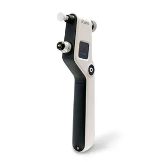
iCare Tonometer
This accurate painless puffless tonometer measures the eye's intra-ocular pressure (IOP).
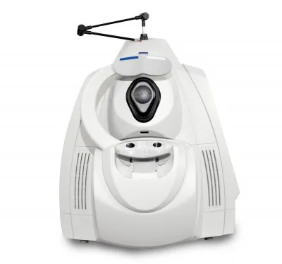
Cirrus 4000
The ZEISS Cirrus HD-OCT 4000 (Cirrus HD-OCT or Cirrus) enable examination of the posterior and anterior of the eye at an extremely fine spatial scale, without surgical biopsy or even any contact with the eye. The Cirrus HD-OCT builds on and refines the retinal imaging technology first introduced with the ZEISS Stratus OCT™. HD-OCT stands for "high-definition optical coherence tomography." Employing the advanced imaging technology of spectral domain optical coherence tomography, Cirrus HD-OCT acquires OCT data about 70 times faster (27,000 vs. 400 A-scans per second) and with better resolution (5 μm vs. ~10 μm axial resolution in tissue), compared to first-generation OCT technology. Cirrus acquires whole cubes of OCT image data, composed of hundreds of line scans, in about the same time as Stratus acquires a six-line scan. You can view these data cubes in three planes, or through three dimensions, giving you access to an extensive amount of retinal image data in one scan. Intended Use The Cirrus HD-OCT with Retinal Nerve Fiber Layer (RNFL), Macular, Optic Nerve Head, and Ganglion Cell Normative Databases is indicated for in-vivo viewing, axial cross-sectional, and three-dimensional imaging and measurement of anterior and posterior ocular structures. Indications for Use
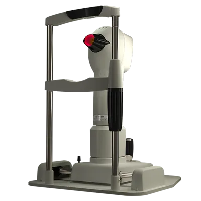
Medmont E300 USB
Produces incredible images of the unique contours of your eye. This is great for fitting sclerals. Advanced vision-assessment system that combines topography, wavefront, autorefraction, keratometry, and pupillometry — allowing accurate and reliable analysis of corneal aberration.
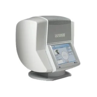
Octopus 600
This is used to assist with Glaucoma detection. It's designed for the examination, analysis and documentation of the field of sight, especially the light difference sensitivity and other functions of the human eye.
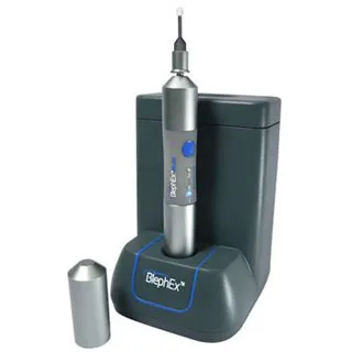
BlephEx
BlephExTM hand piece precisely and carefully, spins a medical grade micro-sponge along the edge of your eyelids and lashes, removing scurf and debris and exfoliating your eyelids.
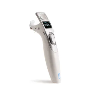
iLux
The iLux® treatment is a gentle, in-office procedure that helps with dry, irritated eyes caused by blocked oil glands (Meibomian Gland Dysfunction). By targeting the source of the problem, it helps your eyes stay healthy and comfortable. Using a small handheld device, your doctor applies soothing heat and gentle pressure to your eyelids to unclog the glands. The treatment is quick, safe, and comfortable, and many patients notice relief soon after.
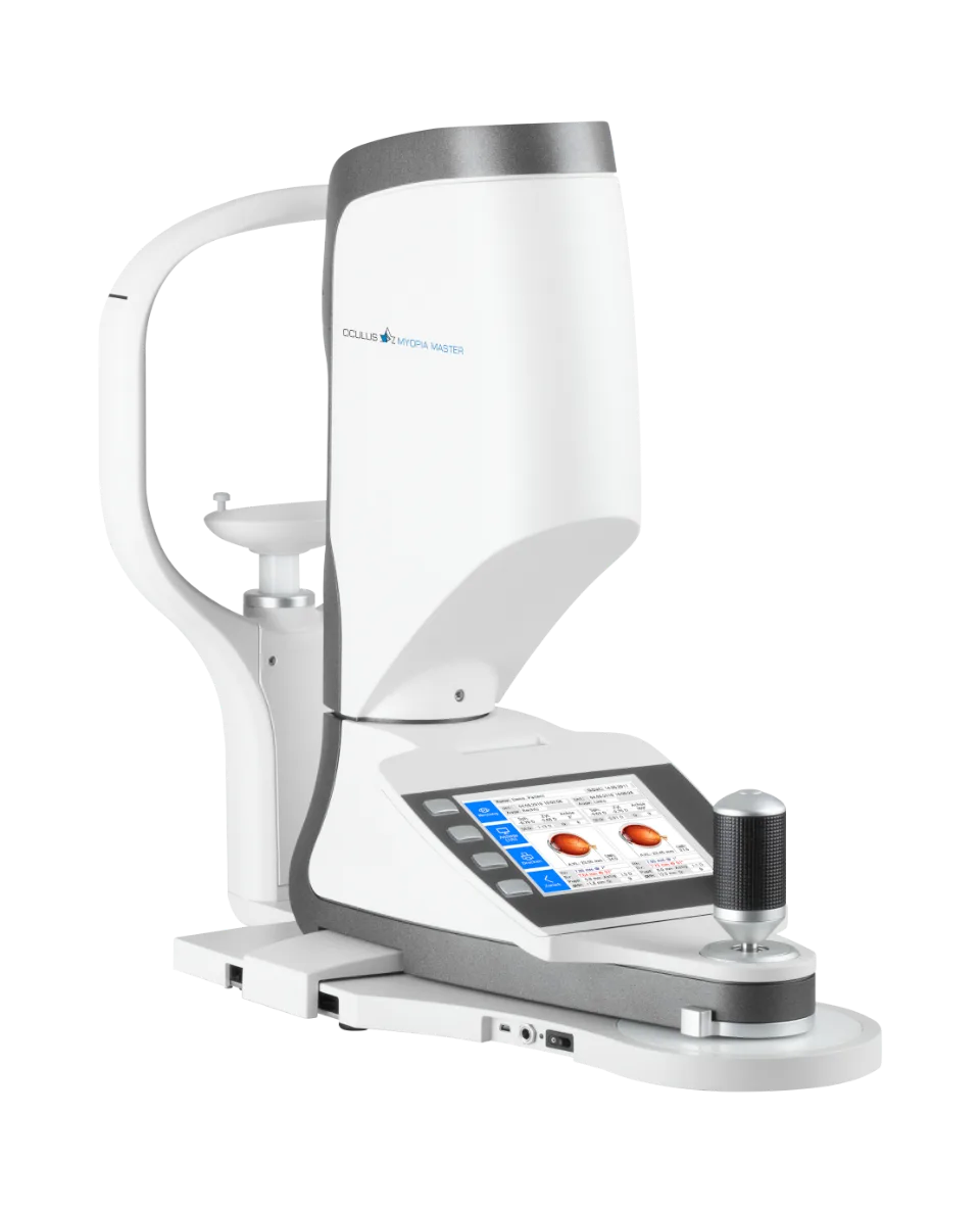
OCULUS Myopia Master®
The OCULUS Myopia Master® is the world’s first device to combine the important measurement methods of myopia management: axial length, refraction values and the central corneal radii. This new machine is regarded by experts as the gold standard for myopia management and a must for every myopia expert. This quick, contactless and extremely accurate measurement method is not influenced by the accommodation-status of the eye and delivers reproducible results also in children. This new technology helps evaluate the effectiveness of reducing the progression of myopia. Please call for more information.